Testing for Glaucoma
Posted by: Eye Consultants of Northern Virginia in Uncategorized

Testing for Glaucoma
Glaucoma is one of the most common eye diseases, affecting about 1.9% of patients over the age of 40. Glaucoma is a slow, chronic disease of the optic nerve that often presents in a very subtle fashion. This disease will affect the peripheral vision first before it causes any loss of visual clarity. Given the slow and chronic nature of this disease, many patients may not be aware of any changes until significant damage has set in. These features make glaucoma a difficult disease to diagnose for both the patient and the physician, which is why we utilize several tests to help determine whether a patient truly has glaucoma and if so, what extent the disease has damaged the optic nerve.
Patients are often referred for glaucoma to the ophthalmologist by their local optometric provider. The normal range for intraocular pressure is about 8-22 mmHg; however, some patients may experience glaucoma even within a “normal range”. An elevated intraocular pressure will often set off a red flag for further investigation into whether a patients is developing glaucoma. Glaucoma is closely tied to the eye’s pressure and the treatment for this disease is linked to lowering the eye pressure. The patient’s cornea, the clear front window of the eye, can lead to variations in the intraocular pressure measurements. One important reading during a glaucoma evaluation is the corneal thickness. When the corneal thickness is thin, patients are at a higher risk of glaucoma as their intraocular pressure readings tend to be falsely low. On the other hand, may patients are referred for elevated intraocular pressure but only have a thick cornea, which has led to falsely elevated eye pressure readings.
Once the eye pressure is taken into account, numerous tests are employed to help evaluate the presence and severity of glaucoma. One important test is Optical Coherence Tomography (OCT). This machine takes a quick picture of the optic nerve and the nerve fibers using reflected light waves, similar to an ultrasound machine. This noninvasive test does not require an “air puff” or any other discomfort to the patient and provides a quantitative measurement to track the disease over time.
Visual field testing is utilized to measure the functional damage from glaucoma. This important test tracks the patient’s performance while detecting subtle peripheral visual field loss. This test does require the patient to press a button when lights flash in the periphery of the vision while they fixate on a central target. This test is critical to help track progression of the disease over time.
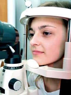
Some other tests for glaucoma include examination under the slit lamp microscope. Gonioscopy utilizes a special lens to visualize the anterior chamber angle. The angle is the area in the corners of the eye where the eye fluid drains out. This network is important to see to determine if any physical obstruction is playing a part in the patient’s glaucoma. Under the microscope, the physician will also examine the optic nerve to determine if there are any other features suggestive of worsening disease over time.
The physician will put together all of these tests to ensure the patient is on the appropriate treatment regimen to help minimize any disease progression.
Drs. Albano, Abramowitz and Van Looveren are glaucoma specialist practicing in both our Woodbridge and Springfield office locations. Contact our office today to schedule your medical consultation.
Posted by Benjamin Abramowitz, MD
